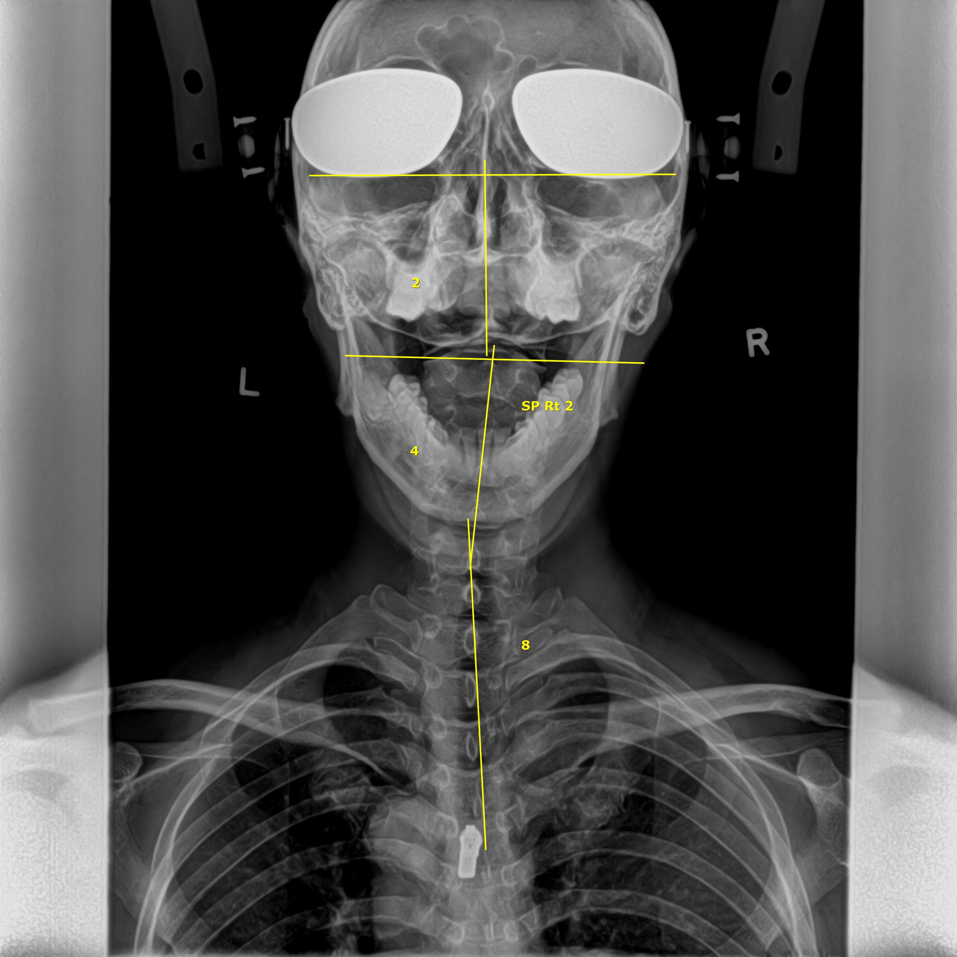
X-rays are a type of electromagnetic radiation most commonly known for their ability to see through a person's skin and reveal images of the bones beneath it. X-rays were first discovered in 1895, where experiments showed that this particular form of radiation could penetrate soft tissues but not bone, resulting in a shadow image that can be projected onto photographic plates. Due to their ability to penetrate certain materials, X-rays are used for several non-destructive evaluation and testing applications, particularly for identifying flaws or cracks in structural components. The radiation beam is directed through a part of the body, or through an object, and onto a film or specialised detector to absorb the X-rays that have penetrated through. The resulting shadowgraph reveals the internal features of the body or object, allowing one to determine whether certain parts are damaged or structurally compromised. This very same application is used by doctors and dentists to create X-ray images of bones and teeth, respectively. With advances in technology, it has led to more powerful and focused X-ray beams, more accurate focusing systems and more sensitive detection methods, such as improved photographic films and electronic imaging sensors. As a result, it is now possible to distinguish, with increasingly fine detail, subtle differences in tissue density; while using much lower amounts of X-rays. It has also led to even greater applications for X-rays like imaging tiny biological cells or structural components of materials like cement to killing cancer cells.
Chiropractic and X-rays
Like how doctors and dentists use X-rays to aid in their diagnosis and treatment plans, chiropractors may also request for X-rays to be done as part of the diagnosis and treatment process. Chiropractors are neuromusculoskeletal specialists, placing special emphasis on the spine, and for good reason too. The spine forms the supporting framework of the body that allows for the movements we have. It also houses the spinal cord, which controls everything from how our muscles move to the proper functioning of our organs. Thus, it is for this reason that chiropractors ensuring the spine is in proper alignment is crucial to our daily activities and our optimal movements. Therefore, the use of X-rays along with using posture pictures, would help chiropractors see the biomechanics of the spine, identify and correcting for any biomechanics issues. Some may argue that the use of posture pictures would suffice and taking X-rays is redundant. Others might say that the chiropractor should be able to feel the alignment issues through proper palpation of the back. While these may be true, they only present half the picture. Surrounding our spine are multiple layers of muscles, which are then enclosed by our skin. Furthermore, the muscles of different individuals vary in size and thickness, which makes it unreliable to identify for misalignments simply through palpation. Importantly, our muscles may contract as a form of guarding mechanisms in response to any misalignments in the spine. As a result, sometimes a posture picture might suggest a mild imbalance in posture and musculature but in reality, the X-ray would reveal a more apparent misalignment in the spine. X-rays would also provide important information that cannot be observed from a posture picture or through palpation. Firstly, X-rays would allow for the visualisation of each individual vertebra of the spine and its associated intervertebral disc. The intervertebral discs are gel like structures that function as shock absorbers. They are well placed between each vertebra of the spine to help absorb and redistribute the load experienced from gravity and our daily activities. An X-ray allows your chiropractor to see how your spine responds to load by requesting for a standing and seated view. The heights of the intervertebral discs can be observed to determine if they are healthy and functioning properly while the position of each vertebrae relative to one another can also be observed to identify for any abnormal movements and biomechanics of the spine. This information would provide an indication as to how your spine is functioning during weight bearing movements, and whether it is able to redistribute the load effectively. Secondly, an X-ray allows your chiropractor to observe the natural curvatures of the spine. A normal healthy spine should have three natural curves – a lordosis (backward-facing curve) in the neck, a kyphosis (forward-facing curve) in the upper back and another lordosis in the lower back. The three natural curves, along with the intervertebral discs allow the spine to act like a spring and efficiently absorb and redistribute forces due to gravity and daily activities such as sitting, standing, walking, running, lifting, climbing, pulling, pushing and so on. However, because of poor habits, such as looking down at our phones or slouching at the computer desk, the natural curves of the spine may be gradually lost. As a result, the spine cannot effectively redistribute load and certain parts of the spine experiences more strain than usual. With an X-ray, your chiropractor can measure the natural curves in your spine, and determine if they are within normal or, have too much or too little curve. Any deviation from the normal would lead to improper load distribution in the spine that would eventually lead to problems like pain, stiffness and even degeneration of the spine over time. Thirdly, an X-ray provides the chiropractor a way to locate for any signs of spinal degeneration, bone spurs or even any pathologies inside our skeletons. Sometimes, due to improper spinal loading from poor postural habits, the spine undergoes degeneration, or the body responds to the increased spinal loading by developing bone spurs to provide more structural stability. Bone spurs are indicative of improper spinal biomechanics and may sometimes encroach onto the intervertebral foramen – an opening for which our spinal nerves branch out from the spinal cord. A foraminal encroachment would therefore potentially cause a nerve impingement that may lead to a multitude of problems like pain, numbness, and organ dysfunction. Therefore, it is important for your chiropractor to identify for any contraindications so they can design the best treatment plan for you. X-rays are essential tools during scoliosis treatment at All Well Scoliosis Centre. Other than the aforementioned details that can be gathered from spinal X-rays, additional important information can be gathered from an X-ray of a scoliosis spine. Scoliosis is a 3-dimensional deformity of the spine and X-rays is a form of mapping of the musculoskeletal functions and biomechanics of our body. Having scoliosis would lead to loss of balance, problems with body coordination and muscles activation. At All Well Scoliosis Centre, the X-rays of scoliosis patients are carefully analysed in detail to design a suitable scoliosis treatment plan specific to your scoliosis conditions. Every scoliosis conditions are different, there is no scoliosis curvature although the shape of the curve are similar on the X-rays, it does not mean will react the same. Thus, the scoliosis treatment at All Well Scoliosis Centre differs between each individuals. Besides measuring the Cobb’s angle to determine the severity of the scoliosis, proper X-rays would also allow your chiropractor to see the amount of rotation the spine has. In cases where there is more than one scoliosis curve, relying on the Cobb’s angle alone without considering spinal rotation would not be sufficient to determine which is the major curve and it may minimal improvement to the condition. In addition, any imbalances in the heights of your hips or shoulders can also be gathered from the X-rays. When looking at an X-ray of a child during growth spurts, the Risser’s sign could be identified, revealing the skeletal maturity of your child. This procedure would help to determine if the scoliosis condition is progressive or not. Any misalignments can also be measured from the X-rays through specific measurements and calculations. The information would then direct the course of your scoliosis treatment when you visit our clinic. Proper X-rays screening enables us to know the proper set up of the rehabilitation devices and proper procedure during active and passive scoliosis treatment exercises. Secondly, we can strategically determine what type of mirror image devices to use and where to place them, along with additional home scoliosis exercises that needs to be done to help correct and re-educate the brain to help you learn the correct posture while performing Sensory Motor Re-integration. Lastly, x-rays would help chiropractor at All Well Scoliosis Centre perform detail spinal manipulation according to the 3-dimensional structure of the spine. By referring to the information on the X-rays, we can determine which area of your spine, hips, or ribs to perform the alignment to help release the accumulated tension in the spine and musculature. Therefore, X-rays are an important part of a chiropractor’s scoliosis treatment procedure. X-rays allow the chiropractor at All Well Scoliosis Centre to see the structure of the spine, allowing them to relate it with your posture and any underlying conditions or complaints you may have. This would then allow them to better tailor the treatment plan to give you the best chances of recovery.
Health concerns of X-rays
It has been widely reported that exposure to radiation from X-rays increases the risk of cancer. While this is true, it is not entirely accurate. Radiation exposure is not what increases our risk of cancer, but rather the dosage of radiation that we are exposed to. High levels of radiation damage the cells in our body and our DNA. This creates errors in our DNA which could accumulate over time, increasing the chances of our cells becoming dysfunctional and developing into cancer. However, the amount of radiation during a routine X-ray is minimal and would not amount too much risk. In fact, we are constantly exposed to natural background radiation every day. Radon gas naturally seeps out from the ground and constitutes 58% of natural background radiation. Other sources of background radiation include those that come from rocks containing natural radioactive elements like uranium, from cosmic rays that stream down onto Earth from the sun, or even from the food we eat – plants can absorb radioactive compounds from the soil and eventually make its way onto our dinner table. The amount of radiation we are exposed to during an X-ray is only equivalent to only a few days of natural background radiation. To put that into perspective, a four-hour air flight will expose the body to the same amount of radiation as an X-ray due to exposure to cosmic rays. Furthermore, during the process of taking an X-ray, every effort is made to minimise the amount of radiation the patient is exposed to. If an X-ray is required that exposes the reproductive system, such as X-rays of the abdomen, lead shields will be placed over them as a precaution and prevention to reduce the amount of radiation exposure. In addition, advancements in X-ray technology has led to more focused X-ray beams and more sensitive detectors that allow for high quality images even at low radiation doses. Therefore, it is evident that the amount of radiation exposure from X-rays is minimal and is not significantly different from what we are exposed to daily, bearing little to no risk to our overall health. In fact, low doses of radiation have been reported to bring some health benefits. Due to the damaging nature of X-rays, low doses of X-rays may help stimulate our body’s own biological protection systems against cellular damage, toxins, and pathogens. This stimulation has been reported to produce a range of beneficial effects by boosting our immune system and even lowering the risk of cancer. With that said, precautions are still needed for certain groups of people like children and pregnant women. As children still have their whole life ahead of them, it is believed that they may be exposed to twice the risk of developing cancer compared to middle-age people for the same X-ray procedure. Thus, X-rays for children are often only requested when it is required for the treatment purposes and the radiation dose will be kept as low as possible. For pregnant women, special precautions are required when an X-ray is performed in a region near the womb. Exposure to radiation could potentially harm the development of the unborn child. However, if the X-ray is essential for diagnosis and treatment, and the health benefits clearly outweighs the small radiation risk, the X-ray will have to be performed and all necessary protections and precautions will be followed.
Conclusion
X-rays are an important diagnostic tool. Although concerns regarding the risk of radiation exposure from X-rays are not unfounded, advancements in X-ray technology has ensured that those risk have become negligible. At All Well Scoliosis Centre, X-rays are an important part of our diagnosis and treatment process. By looking at your 3-Dimensional X-rays, we can properly identify any biomechanics issues with your spine and relate it with your posture and complaints. Regardless if you are a normal or scoliosis patient, having a set of X-rays would reveal important information that would help guide the chiropractor at All Well Scoliosis Centre to determine what kind of chiropractic treatment and/or scoliosis treatment should be provided. From there, we can then carefully tailor a personalised treatment plan to put you on the path of recovery. We fully understand the small risk associated with X-rays and the concerns patients may have. However, we also believe in ensuring our patients feel better following our treatment, and the benefits of providing you with the best chances of recovery clearly outweighs the small risk of radiation exposure from X-rays.
References
1. Oakley PA, Harrison DD, Harrison DE, Haas JW. On “phantom risks” associated with diagnostic ionizing radiation: evidence in support of revising radiography standards and regulations in chiropractic. The Journal of the Canadian Chiropractic Association. 2005 Dec;49(4):264.
2. Oakley PA, Cuttler JM, Harrison DE. X-ray imaging is essential for contemporary chiropractic and manual therapy spinal rehabilitation: radiography increases benefits and reduces risks. Dose-Response. 2018 Jun 18;16(2):1559325818781437
3. X-Rays – Benefits and Risks. Available from: https://www.radiology.ie/images/uploads/2012/01/X-Rays-Benefits-and-Risks.pdf